Hospital + emergency department · APP field guide

Spine Consult

A single, reconciled bedside guide for the 20 highest-yield adult spine presentations encountered on the floor and in the emergency department: triage, localization, imaging, initial management, and escalation.

20condition modules
3urgency tiers
1repeatable consult method
Download print PDF

Andrew J. Pugely, MD, MHCDS
Medical Director - Orthopedics, MercyOne Genesis
Clinical review date: July 19, 2026

Common + degenerative

Most presentations are nonemergent, but every evaluation begins with a red-flag screen.
Condition 01

Acute Nonspecific Neck or Back Pain

Common, usually self-limited - after dangerous alternatives are excluded
Routine / conditional
A normal neurologic examination and absence of red flags support conservative care; “pain intensity” alone does not establish a spine emergency.

Recognize

  • Diffuse axial pain after activity or without a clear event; motion and paraspinal palpation may reproduce symptoms.
  • No objective motor, sensory, reflex, gait, sphincter, constitutional, trauma, cancer, or infection warning pattern.
  • Actively consider nonspinal causes: aortic disease, renal/urinary disease, abdominal or pelvic pathology, hip disease, shingles, cardiopulmonary causes of thoracic pain.

Focused history + exam

  • Document gait and a complete motor-sensory-reflex screen.
  • Ask directly about fever, cancer, immune suppression, osteoporosis/steroids, anticoagulation, recent procedure, urinary symptoms, and minor trauma in older adults.

Initial orders / imaging

  • No routine imaging for uncomplicated acute low-back or cervical pain without red flags.
  • Target testing to the alternative diagnosis when symptoms are atypical. Image earlier for trauma, osteoporosis, cancer, infection risk, significant neurologic findings, or progressive symptoms.

Call spine now when

  • New or progressive motor deficit, myelopathic signs, saddle/bladder change, systemic toxicity, severe thoracic pain with red flags, pulsatile abdominal mass, or pain out of proportion.

First-line management

  • Multimodal nonopioid analgesia when safe; use opioids sparingly with a stop plan.
  • Encourage early activity and avoid prolonged bed rest. Address mobility, sleep, nausea, and constipation barriers.

Disposition

  • Discharge when the examination is stable, pain is manageable, ambulation is safe, and follow-up is reliable.
  • Return immediately for weakness, gait decline, saddle symptoms, urinary retention/incontinence, fever, or escalating unremitting pain.
Pitfall: Ordering MRI for every flare while missing a focused search for nonspinal emergencies.
Condition 02

Lumbar Radiculopathy / Acute Disc Herniation

Root pain is common; progressive weakness or cauda equina physiology is the emergency
Routine → emergent if deficit
Leg pain often exceeds back pain and follows a root pattern; pain severity is less important than motor change, bilateral findings, and sphincter symptoms.

Recognize

  • L4: anterior thigh/medial leg, quadriceps weakness, reduced patellar reflex.
  • L5: lateral leg/dorsal foot, great-toe or ankle dorsiflexion weakness; watch heel walking and foot drop.
  • S1: posterior calf/lateral foot, plantarflexion weakness, reduced Achilles reflex. Cough, sneeze, or Valsalva may worsen pain.

Focused history + exam

  • Grade motor power and distinguish pain inhibition from true weakness. Test heel/toe walking when safe.
  • Straight-leg raise supports L5/S1 irritation; crossed SLR is more specific. Femoral stretch assesses upper lumbar roots.
  • Screen every bilateral or severe presentation for saddle, urinary, bowel, and sexual dysfunction.

Initial orders / imaging

  • No emergent MRI for stable uncomplicated radiculopathy without red flags.
  • MRI without contrast for severe or progressive deficit, suspected CES, or persistent symptoms when intervention is likely. Add contrast for infection, tumor, inflammatory disease, or selected postoperative questions.

Call spine now when

  • Progressive foot drop, quadriceps or plantarflexion weakness, bilateral deficits, saddle symptoms, or bladder/bowel change.

First-line management

  • Analgesia, anti-inflammatory therapy when safe, activity as tolerated, and a focused rehabilitation plan.
  • Do not delay CES imaging for a medication trial.

Disposition

  • Stable pain and examination: outpatient follow-up. Admit for neurologic progression, unsafe mobility, uncontrolled symptoms, or a serious alternative diagnosis.
Pitfall: Calling all leg pain “sciatica”; vascular claudication, hip disease, peripheral neuropathy, and DVT can mimic radiculopathy.
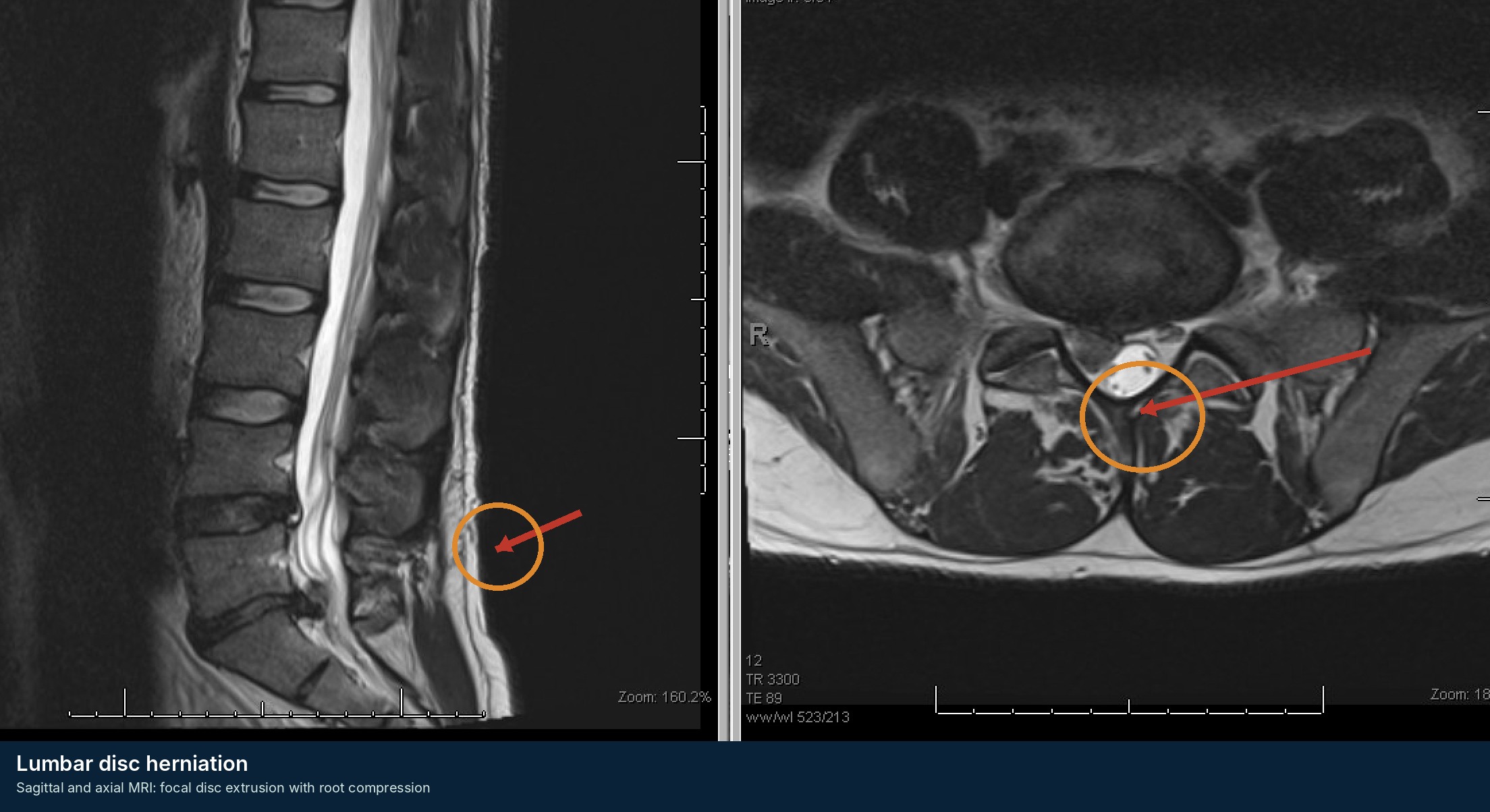
Clinical imaging example for Lumbar Radiculopathy / Acute Disc Herniation
De-identified teaching image from the Andrew J. Pugely, MD educational archive; annotations are schematic and not a substitute for formal interpretation.
Condition 03

Cervical Radiculopathy

Dermatomal arm pain with a lower-motor-neuron root pattern
Routine → urgent if motor deficit
Differentiate a single-root syndrome from cervical myelopathy, shoulder disease, peripheral entrapment neuropathy, and cardiac or vascular causes.

Recognize

  • C5: deltoid weakness; C6: wrist extension/thumb; C7: triceps/middle finger; C8: finger flexion/ulnar hand; T1: finger abduction.
  • Arm pain may worsen with neck extension/rotation or Valsalva; shoulder abduction may relieve root tension.
  • Myelopathic symptoms - gait imbalance, hand clumsiness, hyperreflexia, bowel/bladder change - move the problem out of the “routine radiculopathy” pathway.

Focused history + exam

  • Grade deltoid, biceps, wrist extension, triceps, finger flexion, and interossei; compare reflexes and dermatomal sensation.
  • Spurling may reproduce radicular pain but is not required. Examine shoulder, pulses, and peripheral nerve provocation.
  • Document Hoffmann, clonus, Babinski, gait, and hand dexterity when cord disease is possible.

Initial orders / imaging

  • MRI without contrast for progressive motor deficit, myelopathic findings, severe persistent symptoms, or planned intervention.
  • CT is useful for fracture, ossification, and bony foraminal detail; radiographs are mainly for alignment and follow-up.

Call spine now when

  • Progressive objective weakness, gait decline, upper-motor-neuron signs, bilateral symptoms, trauma, infection/tumor concern, or severe unremitting pain with a dangerous alternative diagnosis.

First-line management

  • Analgesia, activity modification, and therapy when stable; avoid prolonged immobilization.
  • Urgent pathway for progressive weakness or cord features.

Disposition

  • Most stable cases are outpatient. Admission is driven by neurologic change, unsafe function, or a serious competing diagnosis.
Pitfall: Attributing hand weakness to “radiculopathy” when the pattern is actually myelopathy or peripheral nerve disease.
Condition 04

Lumbar Spinal Stenosis / Neurogenic Claudication

Multifactorial canal narrowing with posture-dependent leg symptoms
Routine → urgent if rapid change
Classic neurogenic claudication worsens with standing/walking and improves with sitting or flexion; a static MRI finding is not itself an emergency.

Recognize

  • Bilateral or asymmetric leg heaviness, pain, numbness, or weakness with standing and walking; “shopping-cart sign.”
  • Symptoms often improve with sitting or lumbar flexion and may coexist with spondylolisthesis or degenerative scoliosis.
  • Differentiate vascular claudication, hip/knee arthritis, neuropathy, and deconditioning.

Focused history + exam

  • Document walking tolerance, gait, pulses, hip motion, motor grades, reflexes, and sensory pattern.
  • Ask whether symptoms are chronic baseline or have abruptly changed; new sphincter or saddle findings require the CES pathway.

Initial orders / imaging

  • MRI without contrast when symptoms are disabling, neurologic deficits are present, or intervention is being considered.
  • Standing radiographs can show slip or deformity; CT adds bony detail. Do not equate radiographic stenosis with symptom severity.

Call spine now when

  • Rapidly progressive motor deficit, new inability to walk, saddle/bladder symptoms, acute severe decline after procedure/trauma, or imaging showing high-grade compression with neurologic change.

First-line management

  • Analgesia, mobility aids, therapy, fall-risk reduction, and selective injections or surgical referral in nonemergent pathways.

Disposition

  • Stable chronic symptoms generally do not require admission. Admit for unsafe mobility, acute neurologic decline, or an alternative serious diagnosis.
Pitfall: Using the phrase “severe stenosis” on MRI as a substitute for documenting the patient’s neurologic trend and function.
Condition 05

Degenerative Cervical Myelopathy

Progressive spinal cord dysfunction from cervical degeneration
Urgent → emergent if rapid deficit
Hand clumsiness plus gait imbalance is the high-yield clinical pair; patients may have little neck pain and still have important cord dysfunction.

Recognize

  • Loss of dexterity, dropping objects, handwriting/buttoning difficulty, gait imbalance, falls, leg stiffness, and paresthesias.
  • Upper-motor-neuron signs include hyperreflexia, Hoffmann, inverted brachioradialis, clonus, and Babinski; signs may be incomplete.
  • A stepwise or rapidly progressive decline is more urgent than a long stable baseline.

Focused history + exam

  • Observe gait, tandem gait, grip-release, finger escape, hand intrinsic strength, and lower-extremity tone.
  • Map motor and sensory findings; document baseline function and serial change.
  • Exclude mimics including neuropathy, Parkinsonism, stroke, normal-pressure hydrocephalus, B12 deficiency, and tandem lumbar stenosis.

Initial orders / imaging

  • MRI cervical spine without contrast is the key test. Add CT for ossification, fracture, or bony anatomy and standing radiographs for alignment.
  • Cord signal change supports chronic injury but absence of signal change does not exclude clinically important DCM.

Call spine now when

  • Rapidly progressive weakness, new loss of ambulation, acute decline after trauma, sphincter dysfunction, or severe compression with changing examination.

First-line management

  • Protect from falls and high-risk manipulation; use a collar only when trauma/instability is suspected.
  • Moderate/severe or progressive DCM usually requires surgical evaluation; stable mild disease may follow a monitored pathway under surgeon direction.

Disposition

  • Same-day or expedited spine evaluation for progression. Admit when function is unsafe or the examination is changing.
Pitfall: Treating gait decline as “lumbar stenosis” without examining the hands, reflexes, and cervical cord.
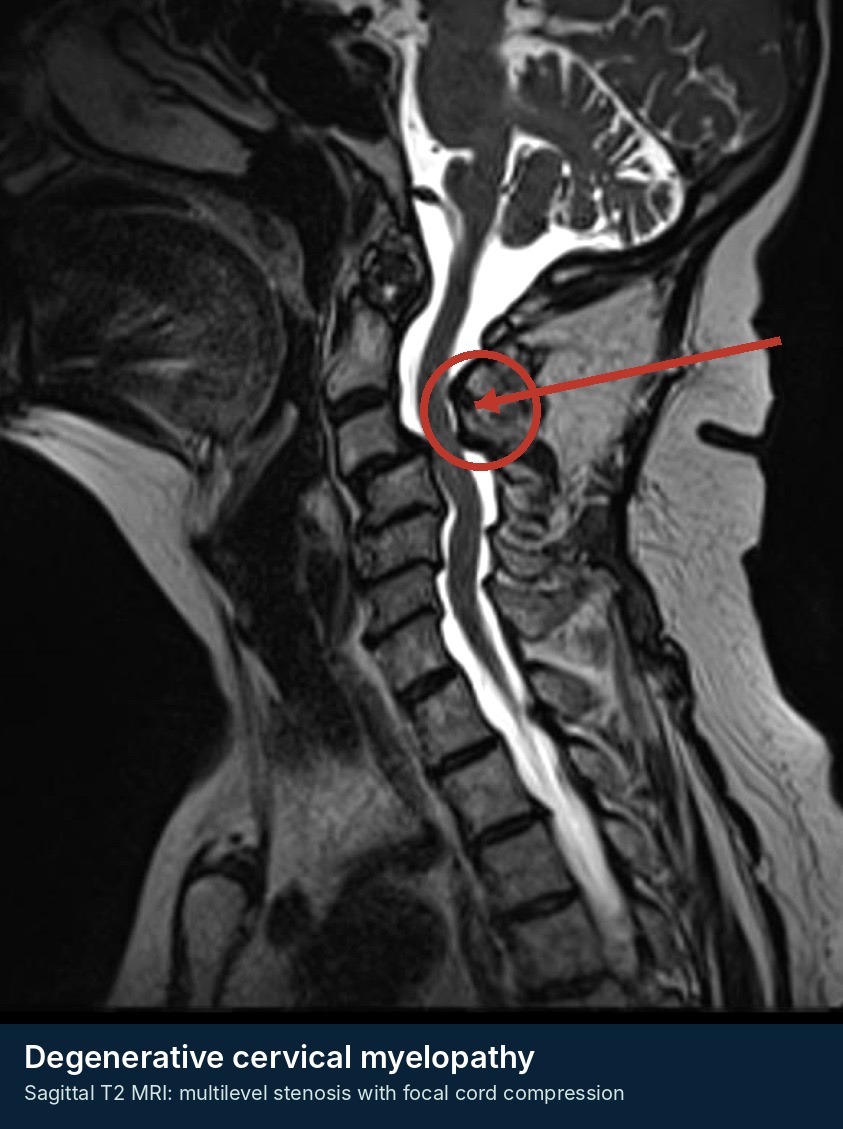
Clinical imaging example for Degenerative Cervical Myelopathy
De-identified teaching image from the Andrew J. Pugely, MD educational archive; annotations are schematic and not a substitute for formal interpretation.
Condition 06

Degenerative Spondylolisthesis / Segmental Instability

A slip is a structural finding; symptoms and neurologic change determine urgency
Routine → urgent if deficit
Most degenerative slips are chronic and low grade. The inpatient question is whether there is acute neurologic compromise, fracture, infection, or inability to mobilize.

Recognize

  • Axial mechanical pain, radiculopathy, or neurogenic claudication; most commonly L4-5.
  • Symptoms may be posture-dependent and coexist with facet arthropathy, stenosis, and degenerative scoliosis.
  • Acute severe pain after trauma or systemic illness should not be assumed to be “just the slip.”

Focused history + exam

  • Document neurologic function, gait, pain with weight bearing, and hip/SI examination.
  • Ask about baseline imaging and chronicity; compare old films when available.

Initial orders / imaging

  • Standing AP/lateral and flexion-extension radiographs are useful for alignment and motion in stable outpatients.
  • MRI answers neural compression; CT answers fracture and bony anatomy. Dynamic films are not for an acutely injured or unstable patient.

Call spine now when

  • Progressive deficit, CES symptoms, destructive change, suspected infection/tumor, acute traumatic instability, or inability to mobilize with concerning imaging.

First-line management

  • Analgesia, activity modification, therapy, and outpatient surgical decision-making when stable.

Disposition

  • Routine outpatient pathway for stable symptoms; admit only for neurologic change, unsafe mobility, or a serious alternative diagnosis.
Pitfall: Letting a familiar chronic slip distract from a new diagnosis above, below, or outside the spine.

Neural emergencies

Sacral, cord, and rapidly compressive syndromes require parallel notification, imaging, and stabilization.
Condition 07

Cauda Equina Syndrome / Conus Medullaris Syndrome

Sacral and multiroot dysfunction - emergency MRI and immediate surgeon notification
Emergent
No single symptom, rectal examination, or post-void residual reliably excludes CES. Call and image in parallel when the pattern is credible.

Recognize

  • New urinary retention, difficulty initiating flow, loss of urinary sensation, reduced stream, overflow incontinence, saddle change, bowel/sexual dysfunction, or bilateral/multiroot symptoms.
  • Conus lesions may be more symmetric with mixed upper- and lower-motor-neuron signs; cauda lesions are often asymmetric and lower-motor-neuron, but overlap is common.
  • Patients may still walk and pain can be severe, mild, or absent.

Focused history + exam

  • Ask specific urinary questions rather than “any incontinence?” Map perineal sensation and document lower-extremity motor/reflex findings.
  • Measure PVR when available, but do not use a low value - including <200 mL - to cancel a concerning history/examination.
  • Rectal tone may add information in selected cases but is not a stand-alone rule-out test.

Initial orders / imaging

  • Emergency MRI of the lumbar spine; extend imaging if the neurologic level is unclear. CT myelography is a specialist-coordinated alternative when MRI is impossible.

Call spine now when

  • Any credible new saddle, bladder, bowel, sexual, bilateral, or progressive multiroot dysfunction.

First-line management

  • Call spine while MRI is arranged; NPO, IV access, serial time-stamped exams, and treatment of hypotension, sepsis, or coagulopathy.
  • Do not delay for analgesic trials, steroids, physical therapy, or an isolated reassuring bedside finding.

Disposition

  • Admit/transfer to a definitive spine center. Operative timing is individualized, but the pathway is immediate.
Pitfall: Waiting for painless retention, complete saddle anesthesia, and bilateral paralysis before acting.
Condition 13

Acute Traumatic Spinal Cord Injury / Central Cord Syndrome

Neurologic resuscitation begins before the MRI
Emergent
Prevent secondary injury: immobilize, avoid hypotension/hypoxia, perform serial standardized examinations, and involve the spine/SCI team immediately.

Recognize

  • Weakness, sensory level, sacral dysfunction, or respiratory compromise after trauma.
  • Central cord classically causes arm/hand weakness greater than leg weakness after hyperextension in an older stenotic spine, often without fracture.
  • Neurogenic shock is hypotension with inappropriate bradycardia after high cord injury; exclude hemorrhage first.

Focused history + exam

  • Document ISNCSCI/ASIA motor and sensory levels and sacral sparing; record last known neurologic baseline.
  • Repeat after transfers, reduction, hypotension, or any clinical change. Assess respiratory function with high cervical lesions.

Initial orders / imaging

  • CT for fracture/alignment; MRI for cord edema/hemorrhage, disc/ligament injury, and persistent compression.

Call spine now when

  • Any suspected cord injury or central cord syndrome - do not wait for MRI confirmation.

First-line management

  • Airway/oxygenation, motion restriction, temperature/glucose control, DVT/pressure-injury prevention, and institutional SCI perfusion protocol.
  • Current AO Spine/Praxis guidance supports early decompression within 24 hours as an option and suggests a MAP lower limit roughly 75-80 with an upper range 90-95 mmHg for 3-7 days; apply local ICU protocol and patient-specific judgment.
  • Routine high-dose methylprednisolone is not a default emergency treatment.

Disposition

  • ICU/trauma admission or transfer to a definitive SCI center.
Pitfall: Reassurance from “no fracture” in a patient with central cord physiology.
Condition 17

Spinal Epidural Hematoma

Abrupt pain followed by rapidly evolving neurologic deficit - MRI and reversal now
Emergent
Time to decompression and the preoperative neurologic state strongly influence outcome. Call while MRI and reversal are occurring.

Recognize

  • Sudden severe neck/back or radicular pain followed by weakness, sensory loss, gait failure, or bowel/bladder change.
  • Risk contexts: anticoagulation, coagulopathy, thrombocytopenia, vascular malformation, trauma, neuraxial procedure, or recent surgery; spontaneous cases occur.
  • Symptoms can progress over minutes to hours and may fluctuate.

Focused history + exam

  • Record exact onset and serial time-stamped motor/sensory findings. Review all antithrombotics, last dose, renal function, platelets, INR/aPTT, and recent procedures.
  • Assess airway and respiratory function for high cervical lesions.

Initial orders / imaging

  • Emergency MRI of the symptomatic spine; extend if localization is uncertain. CBC, platelets, coagulation studies, and type/screen. CT is less sensitive for the collection.

Call spine now when

  • Any suspected epidural hematoma with neurologic symptoms or imaging compression.

First-line management

  • Stop anticoagulants and obtain immediate agent-specific reversal guidance. NPO, IV access, frequent examinations, and emergent decompression planning.
  • Do not perform lumbar puncture or an epidural injection.

Disposition

  • Admit/transfer emergently. Observation of minimal or improving deficits is specialist-directed only.
Pitfall: Attributing sudden severe pain to spasm because the first neurologic examination is normal.

Trauma + fragility

Classify morphology and stability; protect alignment and repeat the neurologic examination.
Condition 08

Cervical Spine Trauma

Clearance, alignment, fracture morphology, and neurologic protection
Emergent if deficit or unstable injury
The absence of neurologic deficit does not imply stability. Maintain alignment, use validated clearance rules, and let CT define bone and alignment.

Recognize

  • Neck pain, tenderness, deformity, distracting injury, altered mental status, intoxication, or neurologic symptoms after trauma.
  • Older adults can sustain odontoid or subaxial fractures after ground-level falls. Ankylosed spines fail with low energy.
  • Facet dislocation/translation, widened interspinous spaces, malalignment, and neurologic deficit suggest instability.

Focused history + exam

  • ABCs first. Preserve pre-injury alignment; document a complete motor-sensory examination before and after transfers.
  • In alert stable patients, NEXUS or the Canadian C-spine rule can identify selected patients who do not need imaging; apply the rule exactly.
  • Perform a formal ISNCSCI/ASIA examination when spinal cord injury is suspected.

Initial orders / imaging

  • CT without contrast is first-line when imaging is indicated.
  • MRI for neurologic deficit not explained by CT, suspected cord/ligament/disc injury, epidural hematoma, or selected obtunded patients under protocol.
  • CTA neck when fracture pattern or mechanism raises vertebral/carotid injury concern.

Call spine now when

  • Any deficit, fracture/dislocation, malalignment, suspected ligament injury, ankylosed-spine trauma, persistent severe pain with concerning mechanism, or inability to clear the spine.

First-line management

  • Rigid motion restriction, log-roll/transfer precautions, NPO, serial exams, and hemodynamic support. Do not force a kyphotic patient into neutral.

Disposition

  • Admit or transfer unstable injuries and all cord injuries. Stable fracture treatment, collar type, and follow-up are surgeon-directed.
Pitfall: Removing a collar because the patient is neurologically intact or a plain radiograph is “negative.”
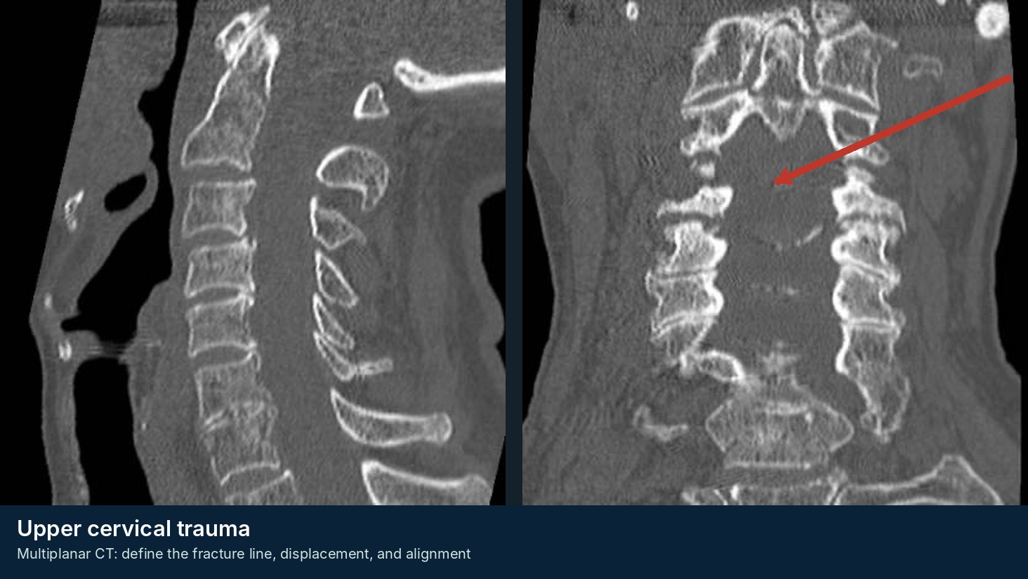
Clinical imaging example for Cervical Spine Trauma
De-identified teaching image from the Andrew J. Pugely, MD educational archive; annotations are schematic and not a substitute for formal interpretation.
Condition 09

Thoracic and Lumbar Spine Trauma

Morphology + posterior ligamentous complex + neurologic status drive stability
Emergent if deficit or unstable injury
CT defines fracture morphology; MRI answers cord, conus, epidural, disc, and ligament questions. The neurologic trend matters more than canal compromise alone.

Recognize

  • Compression fractures are often stable; burst fractures involve the posterior vertebral body wall; flexion-distraction and translational injuries are usually unstable.
  • Thoracolumbar junction injuries are common because of the transition from rigid kyphosis to mobile lordosis.
  • Assess for chest, abdominal, pelvic, calcaneal, and other high-energy associated injuries.

Focused history + exam

  • ABCs and serial motor-sensory-reflex examination, including sacral function when conus/cauda injury is possible.
  • Inspect for a seat-belt sign, palpable gap, deformity, or severe focal tenderness.

Initial orders / imaging

  • CT with sagittal/coronal reconstructions is the principal test.
  • MRI for deficit, suspected posterior ligamentous complex injury, epidural hematoma, occult injury, or discordance between examination and CT.
  • TLICS/AO concepts help communication but do not replace surgeon judgment.

Call spine now when

  • Any neurologic deficit, suspected unstable morphology, progressive deformity, significant canal compromise with symptoms, open injury, or inability to mobilize.

First-line management

  • Spinal precautions, NPO, analgesia, DVT prevention when admitted, and avoidance of hypotension. Brace and mobilization plans are surgeon-directed.

Disposition

  • Admit/transfer unstable injuries, deficits, uncontrolled pain, or unsafe mobility. Selected stable fractures may discharge with a defined brace/activity/follow-up plan.
Pitfall: Assuming retropulsion equals mandatory surgery - or assuming intact neurology equals stability.
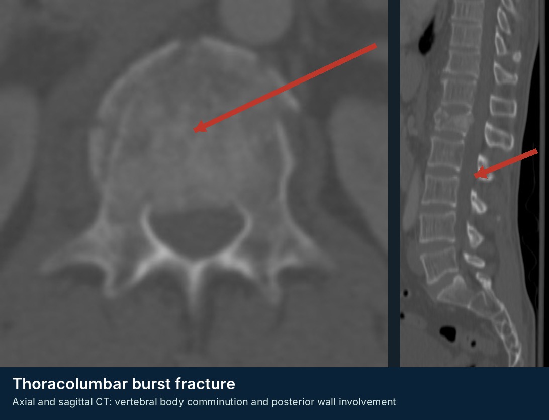
Clinical imaging example for Thoracic and Lumbar Spine Trauma
De-identified teaching image from the Andrew J. Pugely, MD educational archive; annotations are schematic and not a substitute for formal interpretation.
Condition 10

Osteoporotic Vertebral Compression Fracture

A fragility fracture is both a pain problem and a sentinel osteoporosis event
Routine → urgent if deficit/instability
Most simple compression fractures are neurologically intact and treated nonoperatively, but posterior wall involvement, atypical imaging, or severe functional decline changes the pathway.

Recognize

  • Acute focal thoracic or lumbar pain after low-energy trauma, cough, or no clear event in an osteoporotic patient.
  • Pain is often mechanical and worse upright; neurologic deficit is uncommon in a simple wedge fracture.
  • Consider malignancy, infection, burst fracture, and ankylosed-spine injury when the pattern is atypical.

Focused history + exam

  • Percussion tenderness, kyphosis, complete neurologic examination, gait/transfer safety, and hip/pelvic assessment.
  • Review osteoporosis risks: prior fragility fracture, steroids, low body weight, smoking, and treatment history.

Initial orders / imaging

  • Radiographs may identify a wedge deformity but cannot reliably date it.
  • MRI shows marrow edema, neural compression, and malignant/infectious features; CT defines posterior wall and fracture morphology.

Call spine now when

  • Neurologic deficit, burst morphology, progressive collapse, suspected malignancy/infection, ankylosed spine, or pain/immobility refractory to initial treatment.

First-line management

  • Analgesia, early mobilization, selective bracing, fall prevention, and osteoporosis treatment referral. Avoid prolonged bed rest.
  • Vertebral augmentation is selective, not an automatic response to every acute fracture.

Disposition

  • Discharge if pain and mobility are safe. Admit for uncontrolled pain, inability to transfer, medical complexity, or concerning morphology.
Pitfall: Treating the fracture but never initiating secondary fracture prevention.
Condition 11

Sacral Insufficiency / Occult Sacral Fracture

Frequently missed because the sacrum is incompletely visualized on routine lumbar studies
Routine → urgent if neurologic/displaced
In an older osteoporotic patient with severe buttock/groin pain and negative lumbar imaging, image the sacrum and pelvis deliberately.

Recognize

  • Low-back, buttock, sacral, or groin pain after minor trauma or no clear event; worse with weight bearing.
  • Risk factors include osteoporosis, pelvic radiation, steroids, and frailty. Bilateral vertical fractures may form an H pattern.
  • Neurologic deficits are uncommon in insufficiency fractures but can occur with displaced traumatic/U-type injuries.

Focused history + exam

  • Palpate sacrum and pelvic ring; assess hip motion and ability to transfer/walk.
  • Document lower-extremity and bowel/bladder function when pain is severe or the pattern is atypical.

Initial orders / imaging

  • CT pelvis/sacrum defines cortical fracture and ring anatomy but can miss early insufficiency injury.
  • MRI pelvis/sacrum is most sensitive for marrow edema and occult fracture. Do not assume a lumbar MRI fully evaluates the sacrum.

Call spine now when

  • Neurologic deficit, displaced U-type fracture/spinopelvic dissociation, high-energy injury, open fracture, or concern for unstable pelvic ring injury.

First-line management

  • Analgesia, walker, protected or weight bearing as directed by stability, therapy, DVT prevention when admitted, and osteoporosis management.

Disposition

  • Stable insufficiency fractures often follow nonoperative pathways; admit for immobility, pain, or medical optimization.
Pitfall: Repeating lumbar imaging while never imaging the sacrum or pelvis.
Condition 12

Fracture in an Ankylosed Spine (AS, DISH, or Long Fusion)

A rigid spine behaves like a long bone - low-energy trauma can create a three-column injury
Emergent
Preserve the patient’s baseline alignment and assume instability until the entire spine is adequately imaged.

Recognize

  • Ankylosing spondylitis, DISH, extensive fusion, or rigid kyphosis plus new pain after even a ground-level fall.
  • Pain can be deceptively mild and initial radiographs frequently miss fractures. Noncontiguous injuries occur.
  • Delayed displacement, epidural hematoma, and secondary neurologic deterioration are major risks.

Focused history + exam

  • Determine baseline kyphosis and neurologic status; repeat examinations after every transfer.
  • Assess pulmonary reserve, frailty, skin risk, and ability to tolerate supine positioning.

Initial orders / imaging

  • CT of the entire spine with multiplanar reconstruction is commonly appropriate.
  • MRI for neurologic deficit, epidural hematoma, ligament injury, or occult fracture concern.

Call spine now when

  • Any new pain after trauma in an ankylosed spine when fracture is not definitively excluded; immediately for a confirmed fracture or neurologic change.

First-line management

  • Maintain pre-injury alignment with customized padding; do not force the neck or trunk flat. NPO, motion restriction, and serial exams.

Disposition

  • Admit/transfer confirmed or suspected unstable injuries. Operative stabilization is common because bracing often cannot control long-lever biomechanics.
Pitfall: Using a “negative” plain film or a normal first neurologic examination to clear the patient.

Infection + tumor

Pain may precede fever or deficit. MRI, cultures, structural assessment, and multidisciplinary escalation are central.
Condition 14

Spinal Epidural Abscess

Back pain + risk context + inflammatory signal or neurologic change warrants a low threshold for MRI
Emergent
The classic triad of pain, fever, and deficit is usually incomplete at presentation. Diagnostic delay is common and neurologic deterioration can be rapid.

Recognize

  • Severe focal pain, radicular pain, weakness, gait decline, sphincter change, or sepsis.
  • Risk factors: recent S. aureus bacteremia, diabetes, dialysis, immune suppression, injection drug use, indwelling lines, recent injection/procedure, or spine surgery.
  • Fever and leukocytosis may be absent. Cervical/thoracic lesions can threaten the cord and airway/respiration.

Focused history + exam

  • Complete and repeat neurologic examination; document gait if safe. Inspect skin, wounds, lines, and injection sites.
  • ESR/CRP are sensitive screening/follow-up markers but not specific.

Initial orders / imaging

  • MRI with and without contrast of the symptomatic region; consider whole-spine MRI for multifocal symptoms, unclear level, bacteremia, or skip-lesion concern.
  • CBC/CMP, ESR/CRP, and at least two sets of blood cultures. CT-guided sampling may help if cultures are negative and the patient is stable.

Call spine now when

  • Any neurologic symptom, cord/cauda compression, sepsis, rapid progression, cervical disease, or suspected instability.

First-line management

  • Call spine while imaging is arranged. Obtain cultures, then start empiric antibiotics immediately in sepsis, instability, or neurologic compromise.
  • In a stable patient without deficit, coordinate biopsy/culture strategy before antibiotics when feasible. NPO and correct coagulopathy if surgery may be needed.

Disposition

  • Admit. Surgery is common for deficit, compression, instability, failure of medical treatment, or uncontrolled infection.
Pitfall: Waiting for the full triad or repeatedly labeling the patient “back strain.”
Clinical imaging example for Spinal Epidural Abscess
De-identified teaching image from the Andrew J. Pugely, MD educational archive; annotations are schematic and not a substitute for formal interpretation.
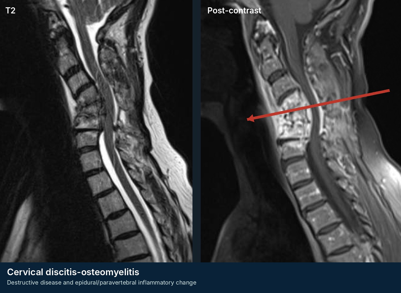
Condition 15

Discitis / Native Vertebral Osteomyelitis

Persistent focal pain and inflammatory markers; microbiology guides treatment
Urgent → emergent if deficit
Stable patients often need cultures/biopsy before antibiotics; unstable, septic, or neurologically compromised patients need immediate empiric treatment and surgical escalation.

Recognize

  • Subacute or progressive focal neck/back pain, often unrelenting and poorly responsive to conservative care.
  • Fever is variable. Bacteremia, dialysis, diabetes, immune suppression, injection drug use, recent infection, or procedure increase risk.
  • May coexist with epidural, psoas, or paraspinal abscess and can produce deformity/instability.

Focused history + exam

  • Serial neurologic examination, focal percussion tenderness, wound/skin/line assessment, and search for a source such as endocarditis, urinary, or skin infection.
  • Review prior blood cultures and recent antibiotics.

Initial orders / imaging

  • MRI with and without contrast; CBC/CMP, ESR/CRP, and two sets of blood cultures.
  • If stable and cultures are nondiagnostic, image-guided biopsy before antibiotics is often preferred. CT defines destruction and guides biopsy.

Call spine now when

  • Neurologic deficit, epidural compression, sepsis, progressive deformity/instability, intractable pain with structural failure, or failure of appropriate antimicrobial treatment.

First-line management

  • Coordinate spine and infectious disease care. Organism-directed prolonged therapy is typical. Brace selectively for pain or instability.
  • Do not use routine follow-up MRI alone to define treatment failure when the patient is clinically improving.

Disposition

  • Admit for sepsis, deficit, uncontrolled pain, instability, or inability to complete a safe diagnostic/treatment plan.
Pitfall: Interpreting persistent MRI enhancement as failure despite improving symptoms and inflammatory markers.
Condition 16

Metastatic Epidural Spinal Cord Compression / Pathologic Fracture

Compression, mechanical instability, tumor biology, systemic status, and goals of care must be assessed together
Emergent if deficit
Pretreatment ambulation predicts outcome. A weak but still ambulatory patient is not a routine oncology follow-up.

Recognize

  • Known or occult cancer with new focal spine pain, night/rest pain, mechanical pain with movement, radicular symptoms, weakness, gait decline, or sphincter dysfunction.
  • Mechanical pain suggests instability even when the neurologic examination is intact. Multifocal disease is common.
  • Common primary tumors include breast, prostate, lung, renal, thyroid, and myeloma.

Focused history + exam

  • Document ambulatory status, exact motor deficits, sensory level, and sacral function.
  • Review histology, disease burden, prior radiation, prognosis, anticoagulation, performance status, and patient goals. Communicate SINS components rather than “looks unstable.”

Initial orders / imaging

  • Urgent MRI of the involved region; whole-spine MRI is often appropriate. CT defines collapse, alignment, and surgical anatomy.
  • Obtain oncology-relevant labs. Avoid unnecessary biopsy when known pathology already establishes the diagnosis and tissue is not needed.

Call spine now when

  • Any objective deficit, high-grade compression, unstable pathologic fracture, rapidly progressive pain, or sphincter dysfunction.

First-line management

  • Call spine, radiation oncology, and medical oncology early. Use dexamethasone per local MSCC protocol after reviewing infection, diabetes, GI, and delirium risks.
  • NPO and spinal precautions when instability or surgery is possible.

Disposition

  • Admit. Treatment may include decompression/stabilization plus radiation, radiation alone, systemic therapy, or comfort-focused care.
Pitfall: Sending a deteriorating ambulatory patient to routine outpatient oncology follow-up.

Postoperative

The operating surgeon should hear about new airway, neurologic, wound, or CSF concerns early.
Condition 18

Postoperative Spine Infection / Wound Problem

The wound, airway, neurologic status, and timing determine urgency
Urgent → emergent if sepsis/deficit/airway
Early postoperative inflammatory changes can be normal; purulence, progressive drainage, dehiscence, systemic toxicity, deep collection, hardware exposure, or neurologic change are not.

Recognize

  • Increasing incisional pain, erythema, warmth, drainage, dehiscence, fever, malaise, or bacteremia.
  • Deep infection may present with pain and elevated CRP without dramatic skin findings. Delayed infection can present with chronic drainage, implant loosening, or pseudarthrosis.
  • After anterior cervical surgery, neck swelling, dysphagia, stridor, or respiratory distress is an airway emergency until proven otherwise.

Focused history + exam

  • Identify operation/date/levels/surgeon, baseline deficit, drain/durotomy, anticoagulation restart, and wound course.
  • Inspect without deep probing; document drainage amount/character, fluctuance, airway/voice/swallowing, and complete neurologic examination.

Initial orders / imaging

  • CBC/CMP, CRP (trend is more useful than a single value), ESR, and blood cultures if febrile/systemically ill.
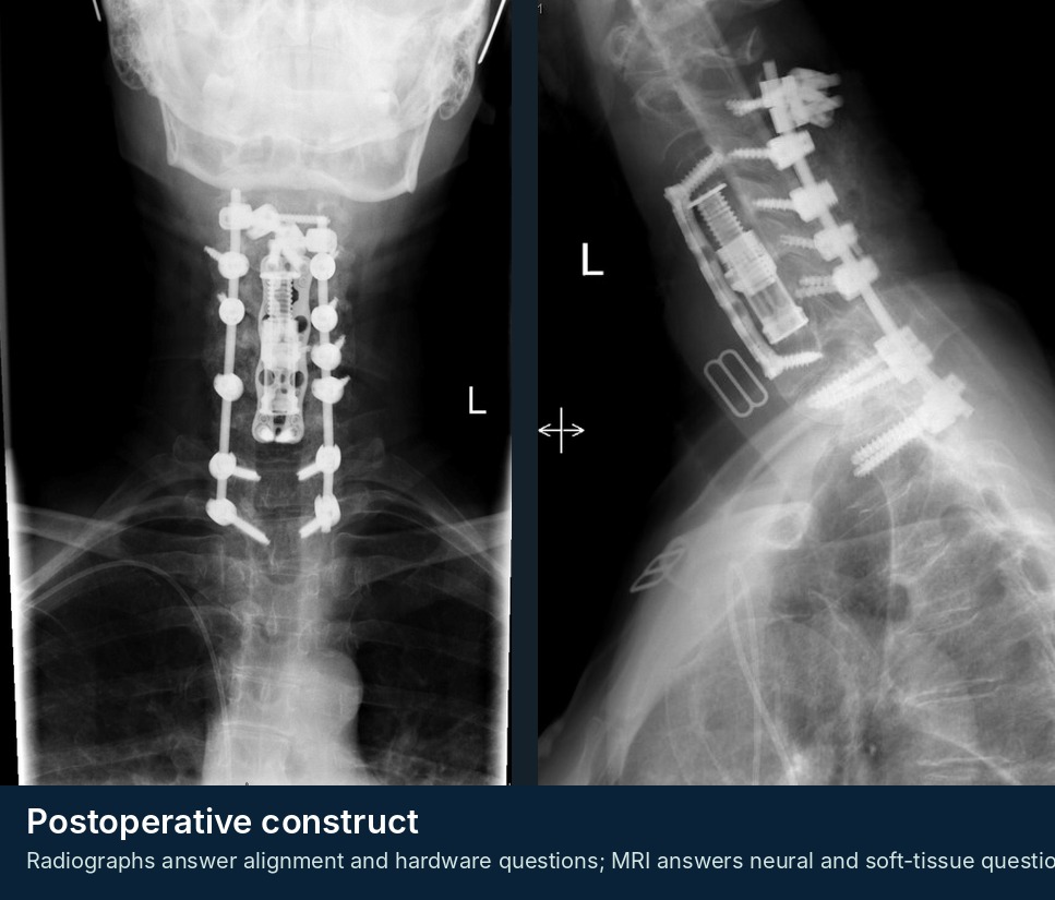
  • MRI with and without contrast for deep collection/neural compression; CT for hardware, fusion, gas, and bony anatomy. Superficial swabs are often misleading.

Call spine now when

  • Airway symptoms, neurologic deficit, sepsis, purulence, dehiscence, exposed hardware, rapidly increasing drainage, or deep compressive collection.

First-line management

  • Notify the operating surgeon. Cultures before antibiotics when stable; immediate antibiotics after cultures in sepsis or neurologic compromise. NPO when debridement is possible.

Disposition

  • Admit for systemic illness, deep infection, airway concern, neurologic change, or need for operative management.
Pitfall: Reassuring yourself with a small skin opening while missing a deep fascial infection.
Clinical imaging example for Postoperative Spine Infection / Wound Problem
De-identified teaching image from the Andrew J. Pugely, MD educational archive; annotations are schematic and not a substitute for formal interpretation.
Condition 19

Acute Postoperative Neurologic Decline

A new deficit after spine surgery is a surgical emergency until proved otherwise
Emergent
The differential is time-sensitive: epidural hematoma, malpositioned hardware/graft, residual compression, cord ischemia, vascular event, abscess, or positional peripheral nerve injury.

Recognize

  • New weakness, sensory change, severe escalating pain, saddle/bladder dysfunction, gait failure, or respiratory compromise after surgery.
  • Symptoms can occur in recovery, after mobilization, or after anticoagulation restart. A normal first postoperative examination does not protect against later decline.
  • After anterior cervical surgery, neck swelling with dysphagia/stridor is an airway emergency.

Focused history + exam

  • Call the operating surgeon immediately and document last known normal. Repeat focused motor/sensory examination and inspect wound/drain/airway.
  • Review operation, levels, implants, blood loss, neuromonitoring changes, intraoperative events, and antithrombotic timing.

Initial orders / imaging

  • CT rapidly assesses hardware, graft, fracture, alignment, and canal compromise. MRI assesses hematoma, residual compression, cord edema/ischemia, abscess, and fluid collection.
  • Imaging choice is individualized; do not delay surgeon involvement while waiting for a final report.

Call spine now when

  • Any new postoperative neurologic or airway change - call now.

First-line management

  • NPO, IV access, oxygen/hemodynamic optimization, urgent coagulation review/reversal, serial exams, and OR readiness.
  • Do not attribute the deficit to anesthesia, pain, or effort until structural and neurologic causes are addressed.

Disposition

  • ICU/OR pathway or transfer to the operating surgeon’s center.
Pitfall: Sending the patient to routine imaging without first activating the surgeon and emergency pathway.
Condition 20

CSF Leak / Pseudomeningocele / Post-Dural Puncture Headache

Positional symptoms and clear drainage require wound assessment and surgeon-directed management
Urgent → emergent if infection/deficit/airway
The urgency is driven by wound failure, meningitis/sepsis, neurologic compression, high-output drainage, intracranial hypotension, and cervical airway concerns.

Recognize

  • Orthostatic headache relieved by lying flat, nausea, photophobia, tinnitus, clear wound drainage, or a fluctuant postoperative collection.
  • Pseudomeningocele may be asymptomatic; complications include nerve-root entrapment, wound breakdown, meningitis, and intracranial hypotension.
  • After cervical surgery, dysphagia or neck swelling has multiple dangerous causes and needs immediate evaluation.

Focused history + exam

  • Identify known durotomy/repair, drain use, timing, wound course, and positionality. Inspect without probing.
  • Document neurologic examination, fever/meningismus, drainage character/volume, and airway/swallowing.

Initial orders / imaging

  • MRI with and without contrast for persistent collection, infection, or neural compression. CT myelography/dynamic studies are specialist-directed for occult leaks.
  • CBC/CRP and cultures when infection is suspected. Beta-2 transferrin is selective when the diagnosis is uncertain and the result will change management.

Call spine now when

  • Neurologic deficit, meningitis/sepsis, wound dehiscence, exposed hardware, high-output persistent drainage, severe intracranial hypotension, airway symptoms, or expanding compressive collection.

First-line management

  • Notify the operating surgeon. Sterile occlusive dressing and short-term positional measures for comfort while the plan is established.
  • Do not repeatedly express or aspirate the collection. Bed rest, drains, blood patch, or reoperation are individualized; antibiotics do not replace source control.

Disposition

  • Small contained leaks may resolve; persistent symptomatic leaks often require targeted intervention. Admit for infection, deficit, wound failure, or inability to maintain hydration/function.
Pitfall: Treating a postoperative orthostatic headache as migraine without examining the wound.