Ten procedures cover most of what walks through this door — from a same-day microdiscectomy to a full deformity reconstruction. Each one below in plain language, with hand-picked video education and trusted resources.
Why this library exists: published research has repeatedly found that most spine-surgery videos online are low quality and produced by unreliable sources. The videos below are independently produced patient education, selected for accuracy and clarity — so you don't have to gamble with a search bar. They illustrate general techniques; your operation is planned on your anatomy, and may differ.
The problem it solves: a herniated disc or bone spur in the neck pressing on a nerve or the spinal cord — arm pain, numbness, weakness, or clumsiness in the hands.
How it works: through a small incision at the front of the neck, the damaged disc is removed, the nerves are decompressed, and a spacer with bone graft is placed so the two vertebrae fuse into one. Because the approach slides between natural tissue planes, recovery is often faster than patients expect.
The problem it solves: the same nerve compression an ACDF treats — but instead of fusing the level, the damaged disc is replaced with an artificial one that keeps the segment moving.
Why it matters: preserving motion may reduce stress on the discs above and below. It isn't right for every neck — bone quality, alignment, and arthritis all factor in — which is exactly the judgment call an experienced surgeon exists to make.
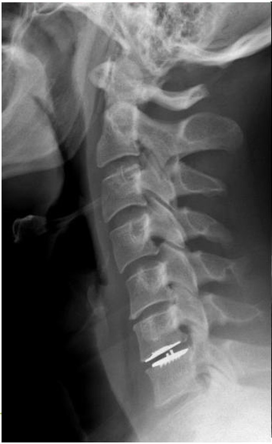
The problem it solves: spinal cord compression across multiple levels (myelopathy), instability, or reconstruction after prior surgery — the bigger cervical problems.
How it works: from the back of the neck, bone compressing the spinal cord is removed and the levels are stabilized with small screws and rods. These are the cervical reconstructions shown in our Case Library.
The problem it solves: sciatica — the shooting leg pain of a herniated disc pressing a nerve root. This is Owen's operation-category: the pain that makes the grocery store impossible.
How it works: through an incision often under an inch, using magnification, only the herniated fragment is removed. The nerve is freed; the disc stays. Leg pain relief is often immediate and dramatic.
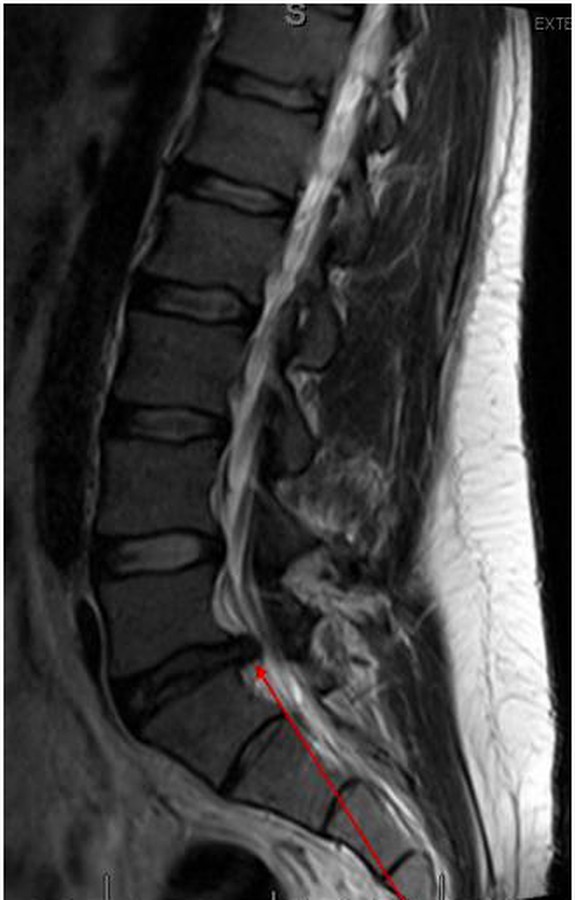
The problem it solves: spinal stenosis — the narrowing that makes legs heavy, numb, and unreliable with walking, classically relieved by leaning on a shopping cart.
How it works: the lamina — bone roofing the spinal canal — and thickened ligament are removed, restoring room for the nerves. Many are done as outpatient surgery, a program our own published research helped establish as safe.
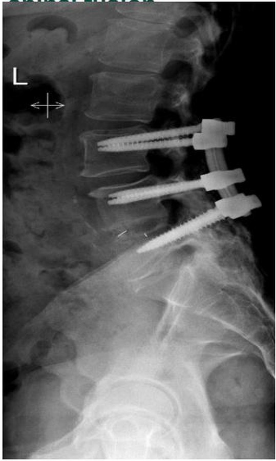
The problem it solves: instability — a slipped vertebra (spondylolisthesis), collapsed disc with nerve compression, or painful degenerative motion that conservative care can't quiet.
How it works: the disc space is cleared and a spacer restores height, decompressing the nerves; screws and rods hold the level while bone grows across it. The approach — from the back (TLIF), front (ALIF), or side — is chosen for your anatomy, increasingly with robotic navigation guiding every screw.
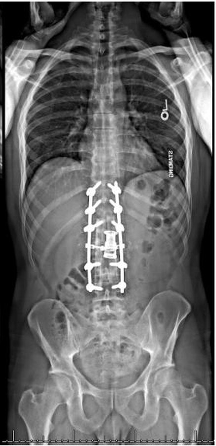
The problem it solves: a spine that has curved or pitched forward — degenerative scoliosis, kyphosis, flatback — leaving you unable to stand straight or walk without leaning.
How it works: full-length standing analysis measures exactly where alignment is lost; correction is planned to the degree, often requiring an osteotomy — precisely reshaping vertebral bone — and long-segment instrumentation, anchored to the pelvis when needed. This is the signature work of this practice, shown film-by-film in the Case Library.
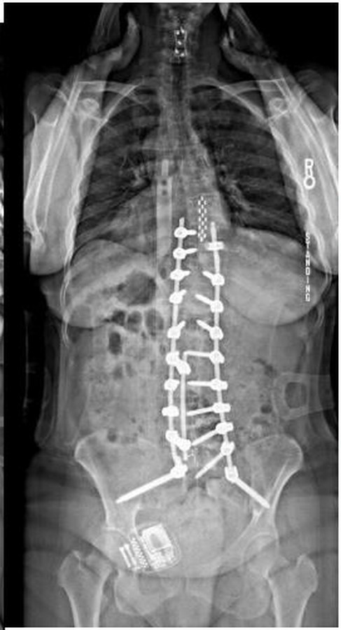
The problem it solves: pain that came back — or never left — after someone else's operation. Failed fusion, broken hardware, adjacent-level breakdown, or alignment that was never addressed.
How it works: there is no standard revision; there is only the forensic workup — imaging that finds what the first surgery missed, then a plan that fixes the actual problem. Owen Shunkwiler's broken hardware was found on exactly this kind of review. Revision is the reason other surgeons have sent their hardest cases here for a decade.
The problem it solves: the sudden, breath-taking back pain of an osteoporotic compression fracture — often from something as small as a cough or a step.
How it works: through a needle, a small balloon gently restores the collapsed vertebra's height; bone cement then stabilizes it from within. Pain relief is often rapid, and the procedure is typically done without an overnight stay.
The problem it solves: sacroiliac joint dysfunction — low back and buttock pain from the joint where the spine meets the pelvis, frequently misdiagnosed as a lumbar problem (and a known cause of pain after lumbar fusion).
How it works: after the diagnosis is confirmed with targeted injections, small implants are placed across the SI joint through a minimally invasive approach, stabilizing it so it can fuse.
If you want to go deeper, go to sources with editorial standards — the same tier of references used in training surgeons.
Ten operations are listed on this page — and the most important decision is often choosing none of them. Surgery is recommended in this practice only when the evidence, your imaging, and your goals all point the same way. If they don't, you'll hear that plainly.
