Iowa Back InstituteLed by Andrew J. Pugely · MD, MHCDS 563-421-4370
The case library — de-identified imaging

The work, on film.

A decade of the state's hardest spine cases — shown the way surgeons see them. Every operation below was performed by Dr. Pugely, start to finish.

All imaging has been de-identified and screened for patient information. These are representative cases illustrating the scope of the practice — every spine is different, and imaging shown does not predict any individual outcome.
New — complete case files

Seven cases, start to finish.

Not just the films — the whole story. Presentation, decision-making, imaging, and outcome for seven de-identified patients. Two of them never needed an operation.

Series 01

Adult deformity & scoliosis reconstruction.

The signature of this practice: full-length standing analysis, alignment measured to the millimeter, and long-segment correction anchored to the pelvis when the case demands it.
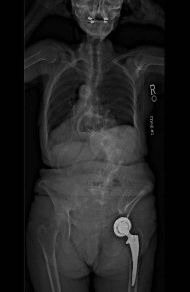
Standing AP X-ray of untreated adult scoliosis
The presentation — untreated adult scoliosis, standing AP
Pre-operative standing AP X-ray showing coronal collapse
One patient, before — coronal collapse, standing AP
Post-operative standing AP X-ray, long construct to the pelvis
Same patient, after — long construct to the pelvis, standing AP
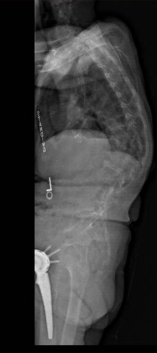
Pre-operative standing lateral X-ray
One patient, before — sagittal alignment, standing lateral
Post-operative standing lateral X-ray, alignment restored
Same patient, after — alignment restored, standing lateral
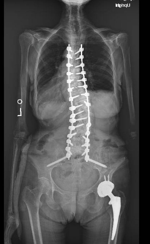
Post-operative standing AP of thoracolumbar-to-pelvis construct
The correction — thoracolumbar-to-pelvis construct, standing AP
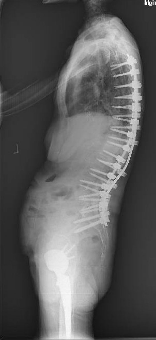
Post-operative standing lateral of long construct
The correction — same case, standing lateral
Post-operative standing AP of long-segment reconstruction
Restored balance — long-segment reconstruction, standing AP
Post-operative standing lateral of long-segment reconstruction
Restored balance — same case, standing lateral
Standing full-length X-rays showing sagittal imbalance
The workup — standing full-length films, sagittal imbalance
Standing X-ray of adult degenerative scoliosis
The presentation — adult degenerative scoliosis
Pre-operative standing AP film of scoliosis
The presentation — coronal deformity, standing AP
Before and after X-rays of thoracolumbar-to-pelvis reconstruction
One patient, before → after — thoracolumbar-to-pelvis reconstruction
Post-operative standing AP of long-segment correction
The correction — long-segment construct, standing AP
Post-operative lateral X-ray with alignment measurements
Measured discipline — lateral with alignment parameters
Post-operative AP of thoracolumbar instrumentation
The correction — thoracolumbar instrumentation, AP
Post-operative lateral of long construct
Restored balance — lateral, long-segment construct
Post-operative AP of long construct
Restored balance — same case, standing AP
Lateral X-ray of kyphosis correction
Kyphosis correction — lateral, post-operative
AP X-ray of long construct with pelvic fixation
Anchored to the pelvis — long construct with pelvic fixation
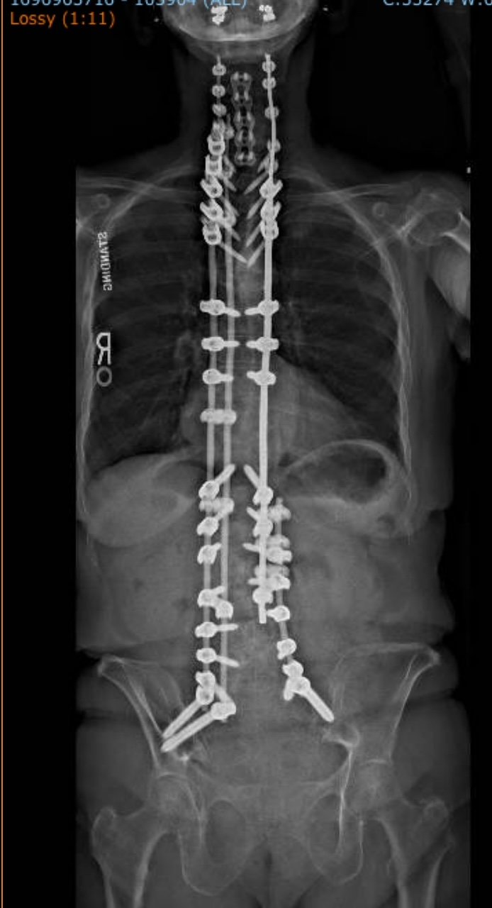
Full-body standing EOS imaging of complex reconstruction
Full-body standing alignment — complex multi-segment reconstruction
Series 02

Complex cervical reconstruction.

Top-5%-in-Iowa volume in cervical spine surgery — from cord decompression to multi-level posterior reconstruction.
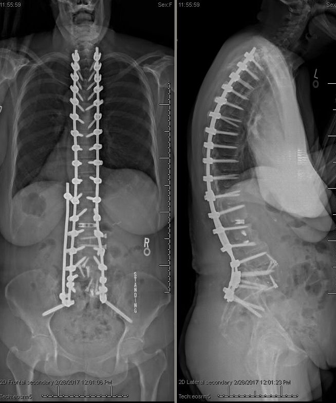
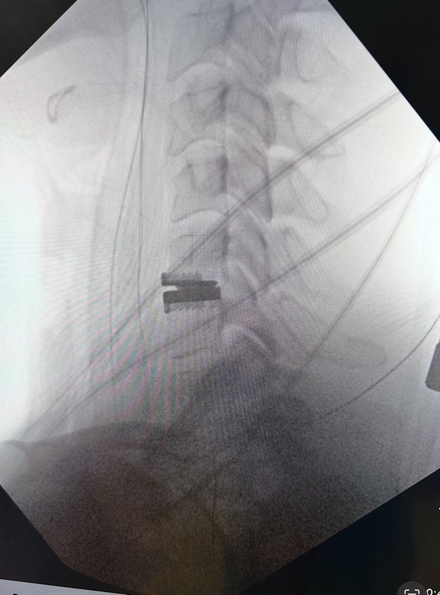
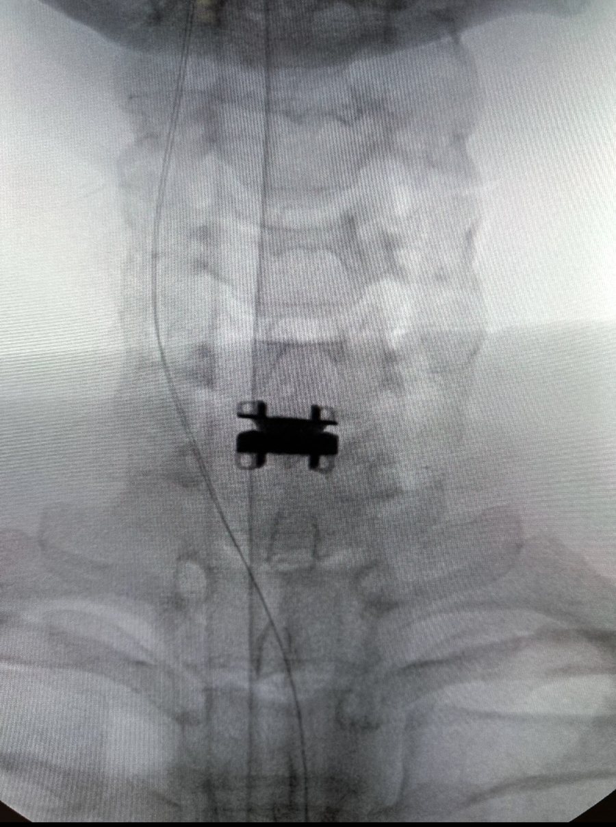
Lateral X-ray of cervical fixation
The construct — cervical fixation, lateral
MRI showing cervical spinal cord compression
The diagnosis — MRI, cervical cord compression
Lateral and AP X-rays of cervical reconstruction
The reconstruction — lateral + AP, posterior instrumentation
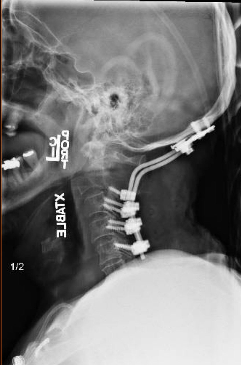
Lateral X-ray of posterior cervical construct
Occiput to thoracic — lateral, posterior construct
AP X-ray of posterior cervical construct
Occiput to thoracic — same case, AP
Series 03

Lumbopelvic & sacral fixation.

When the base of the spine fails — high-grade instability, sacral fracture, failed prior fusion — the fix has to hold where bone quality is worst and forces are highest.

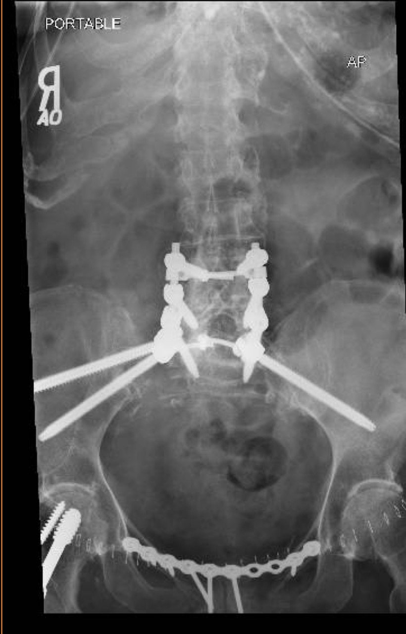
AP pelvis X-ray showing lumbopelvic fixation
The foundation — lumbopelvic fixation, AP pelvis
Sagittal CT of lumbopelvic instrumentation
Verified in section — sagittal CT, lumbopelvic hardware
Series 04

Under the surface.

Advanced imaging drives every decision — where the nerve is pinched, whether the fusion has healed, exactly where the correction must come from.

Lumbar MRI showing spondylolisthesis and stenosis
Where it starts — lumbar MRI, slipped vertebra pressing the nerves
Sagittal CT reconstruction with spinal hardware
Healing, verified — sagittal CT through the construct
Is your case in here somewhere?

Send your imaging. Get a real answer.

563-421-4370
Free imaging review · start online →